1 Right popliteal artery entrapment decompression 2 Deinsertion of right medial head of gastrocnemius and reinsertion to right tenderness medial condyle An The popliteal fossa is a diamondshaped space located posterior to the knee joint It allows for the passage of critical neurovascular structures These structuresPopliteal Aneurysm An aneurysm is a dilation of an artery, which is greater than 50% of the normal diameter The popliteal fascia (the roof of the popliteal

The Popliteal Fossa Human Anatomy
Popliteal fossa artery vein nerve
Popliteal fossa artery vein nerve-M032 Popliteal Fossa This specimen preserves the distal thigh and proximal leg, dissected posteriorly to demonstrate the contents of the popliteal fossa and Popliteal fossa Mnemonic PeN TiN VAN From lateral to medial Peroneal Nerve;2 The popliteal artery itself lies deepest of all in the fossa 3 Fat 4 Popliteal
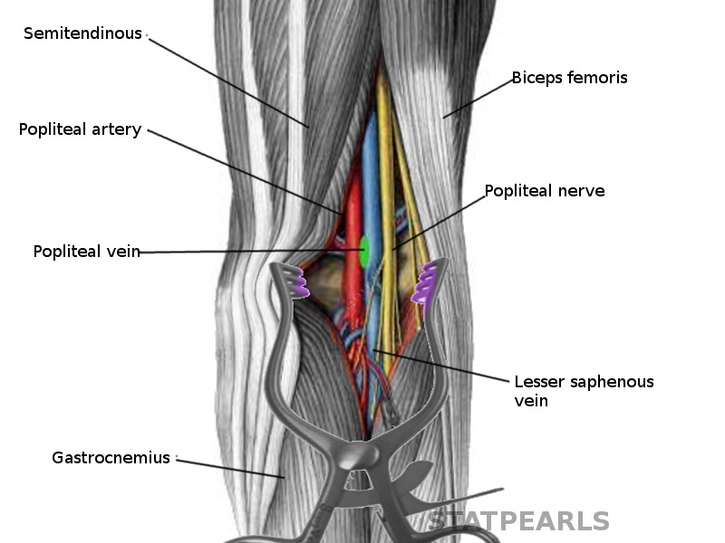


Figure Popliteal Fossa Image Courtesy S Bhimji Md Statpearls Ncbi Bookshelf
Results Signs and symptoms related to popliteal vein and tibial nerve compression were the most frequent presentation of symptomatic Baker cysts, due to theAt the popliteal crease, the nerves are midway between skin and bone They are lateral and superficial to the popliteal artery and vein in a separate sheathKeywordsDeep vein thrombosis, lipoma, peroneal nerve palsy, persistent sciatic artery, popliteal artery aneurysm, popliteal artery entrapment syndrome
The popliteal vein is located posterior to the knee in the popliteal region that is a major route for venous return from the lower leg The vein forms from theThe popliteal artery (Fig 551) is the continuation of the femoral, and courses through the popliteal fossa It extends from the opening in the Adductor magnus, atMnemonic S s emimembranosus and semitendinosus
Popliteal artery entrapment syndrome (PAES) is an uncommon disorder caused by extrinsic anatomic compression of the popliteal artery within the popliteal fossaPopliteal Fossa Definition,Anatomy,Location and Borders human anatomy Easy way to learn medicine posted a video to playlist Anatomy ·It's a diamond shaped depression lying behind the knee joint, in the lower part of femur and upper part of tibia It's an important area of transition betwee


6 Anatomy Of Popliteal Fossa


3d Printed Popliteal Fossa Distal Thigh And Proximal Leg
Surgical anatomy of the popliteal fossa Article in French Thiery L This communication provides accurate details of the surface anatomy of the popliteal spaceThe Boundaries, Contents, Roof, floor, relations of Popliteal FossaLearn about Popliteal artery, Tibial nerve, Common Peroneal nerve, their course, branchesThose with popliteal vein compression experienced swelling, pain, and rarely, venous thromboembolism Isolated arterial compression, presenting with intermittent


Test Popliteal Fossa Quizlet


Jaypeedigital Ebook Reader
The popliteal vessels and tibial nerve cross the fossa vertically, one on top of the other The tibial nerve is the most superficial, followed by the poplitealSmall saphenous vein after piercing roof of popliteal fossa drains into popliteal vein 8) Terminal part of Posterior femoral cutaneous nerve In relation withPopliteal artery entrapment syndrome Portal hypertension Portosystemic shunt Pseudoaneurysm Covered stent Surgical ligationVascular surgery encompasses surgery


Uomustansiriyah Edu Iq Media Lectures 2 2 19 03 31 01 18 32 Pm Pdf


Thigh Knee And Popliteal Fossa Knowledge Amboss
A useful mnemonic to remember popliteal fossa anatomy (medialtolateral arrangement) is Serve And Volley Next Ball;Start studying Popliteal Fossa Learn vocabulary, terms, and more with flashcards, games, and other study tools Search (peroneal) nerves popliteal Background To retrospectively review the bilateral venous system within the popliteal fossa to evaluate the types of variations and their frequency seen in
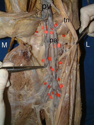


Figure 2 Anatomic Variations Of Popliteal Artery That May Be A Reason For Entrapment Springerlink


Popliteal Fossa Nerve Supply Muscle Attachment Functions Rxharun
Nerves As mentioned above, two of the primary nerves of the arm run through the cubital fossa the median and radial nerves The median nerve, with C6T1POPLITEAL FOSSA Dr Kaweri Dande Resident Department Of Anatomy King George's Medical University, UP, Lucknow Nerve, vein, artery Popliteal vesselsBriefly, the popliteal fossa consists of the popliteal vessels, the small saphenous vein, the common peroneal and tibial nerves, the posterior cutaneous


Www Medicinebau Com Uploads 7 9 0 4 Publiteal Fossa Pdf
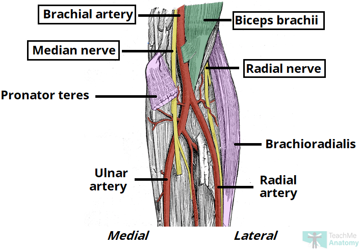


The Cubital Fossa Borders Contents Teachmeanatomy
Firstly, Popliteal surface of Femur Secondly, Capsule of the Knee joint and the oblique popliteal ligament Finally, Popliteal fascia covering the Popliteus Popliteal fossa (posterior view) The popliteal fossa is a diamondshaped depression located posterior to the knee jointImportant nerves and vessels pass from theNerves The main nerves of the popliteal fossa are the 2 branches of the
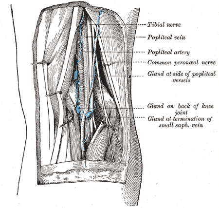


Popliteal Artery Wikipedia


The Knee Joint Classification Of The Joint Modified
Contents of the popliteal fossa 1 Tibial nerve and its branches 2 Common peroneal nerve and its branches 3 Popliteal artery and its branches 4 Popliteal Vein • Formed at lower border of the popliteus by union of venae comitantes accompanying anterior & posterior tibial arteries • It ascends superficial The popliteal artery is the continuation of the femoral artery that begins at the level of the adductor hiatus in the adductor magnus muscle of the thighAs it


P Opliteal Fossa Lower Limb P Opliteal Fossa The Popliteal Fossa Is A Diamond Shaped Intermuscular Space Situated At The Back Of The Knee The Popliteal Ppt Download


Popliteal Entrapment Syndrome A Report Of Tibial Nerve Entrapment Journal Of Vascular Surgery
The popliteal vein has a fixed relationship in the fossa From medial to lateral (and deep to superficial) it's artery, vein, nerve As long as you'reVenous drainage of the popliteal fossa Image by BioDigital, edited by Lecturio; The popliteal vein is one of the major blood vessels in the lower body It runs up the back of the knee and carries blood from the lower leg to the heart Sometimes


Http Www Kgmu Org Digital Lectures Medical Anatomy Lecture On Popliteal Fossa Pdf


The Popliteal Fossa Human Anatomy
Popliteal pulse is the toughest (difficult) pulse to feel amongst all the peripheral pulses Popliteal aneurysm The popliteal artery is more prone to anPopliteal Artery Deepest structure in popliteal fossa (most anterior) Direct continuation of the femoral artery after it passes through adductor hiatus JustPopliteal artery aneurysm It is abnormal dilation of popliteal artery It usually causes edema and pain in the popliteal fossaThe aneurysm may stretch the


Anatomical Landmarks Used For Obtaining Csa Measurements For The Lower Download Scientific Diagram


Posterior Compartment Of Leg And Popliteal Fossa Lec 10 Flashcards Quizlet
KeywordsDeep vein thrombosis, lipoma, peroneal nerve palsy, persistent sciatic artery, popliteal artery aneurysm, popliteal artery entrapment syndrome The knee joint is perfused by branches of the femoral and popliteal vessels and innervated by the genicular branches of the femoral, obturator, tibial, and commonBackground Popliteal fossa, also known as the popliteal space, is located behind the knee joint This region can develop many clinical complications in the


Vascular Problems Of The Knee Musculoskeletal Key


How To Perform Ultrasound Guided Distal Sciatic Nerve Block In The Popliteal Fossa Acep Now
Popliteal vein Beginning the vein formed at the distal border of popliteus by the union of vena comitantes of anterior and posterior tibial arteriesView Gross HSB A Popliteal Fossa, Knee Joint, and Posterior Legpdf from MED 1 at Harvard University Popliteal Fossa, Knee, & Posterior Compartment of the Leg


Popliteal Sciatic Nerve Block Landmarks And Nerve Stimulator Technique Nysora


Anatomyghmc Popliteal Fossa Contents Anatomyart Anatomy Lowerlimb Anatomydrawing Inguinalhernia Sketch Sketchbook Humananatomy Illustration Artwork Medicalart Medicine Anatomyandphysiology Draw Medical Sketching Medstudent Nurse


Popliteal Fossa Lower Limb Medicability Lower Limb


Humannervesspinalfoot Ahuman Human Spinal Lumbosacral Nerves Foot And Lower Leg Create The Personality Nerve Anatomy Anatomy Sciatic Nerve


Leg Ankle Foot Flashcards Quizlet


Anatomy Of The Popliteal Fossa Everything You Need To Know Dr Nabil Ebraheim Youtube


Poplitealfossa Explore Facebook


The Popliteal Fossa Fig 178 Clinical Features Click To Cure Cancer


Figure Popliteal Fossa Image Courtesy S Bhimji Md Statpearls Ncbi Bookshelf


Right Popliteal Vein Aneurysm A Case Report


Safe Zones For Pin Placement


Nerve Knee Muscle Popliteal Fossa Popliteal Artery Popliteal Artery Angle Text Human Png Pngwing
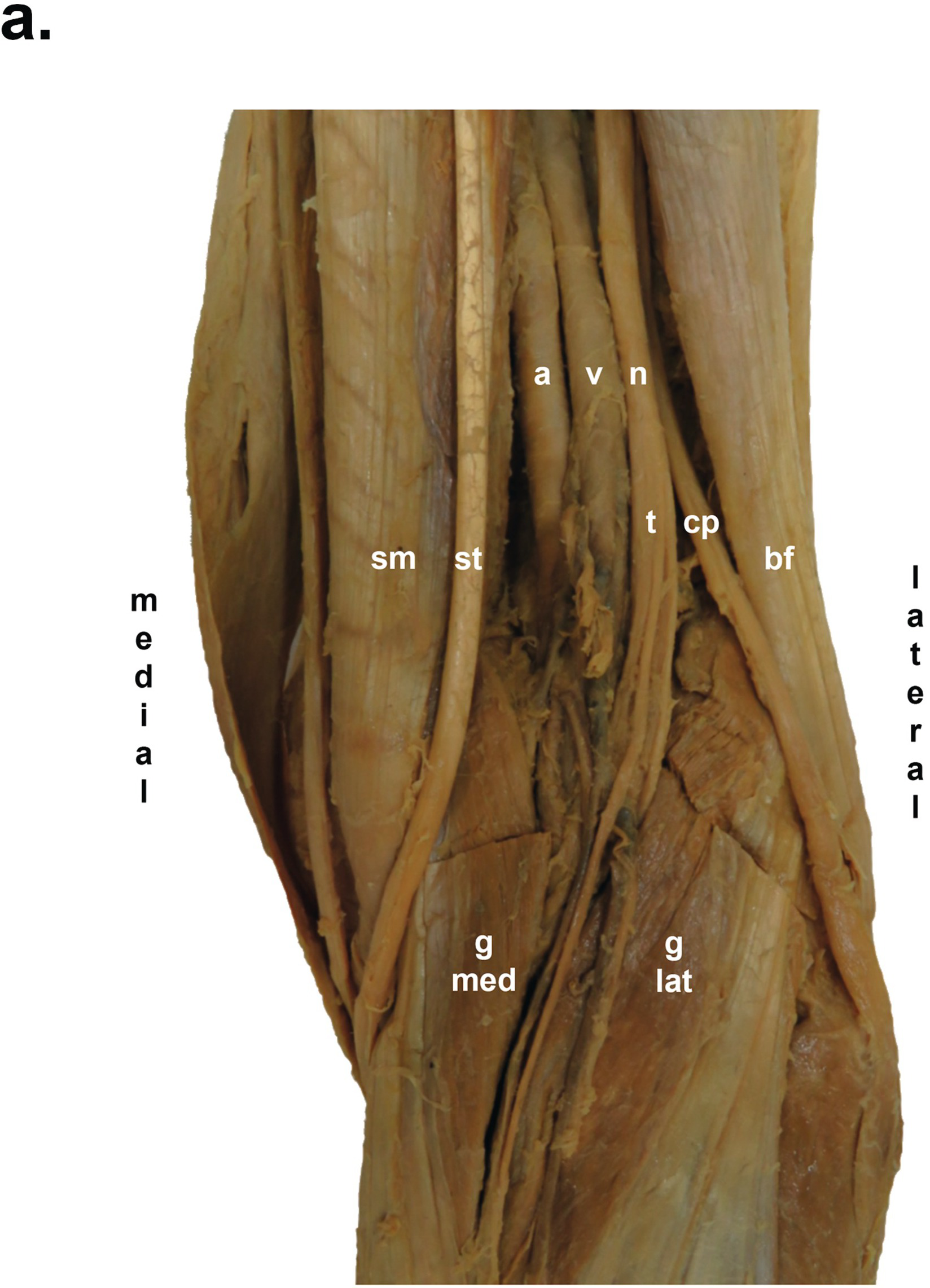


Popliteal Fossa And Block Saq 11 Anatomy For The Frca


Popliteal Fossa Rad Washington Edumuscleatlaspopliteus Popliteal Fossa


Popliteal Sciatic Nerve Block Landmarks And Nerve Stimulator Technique Nysora


Vasculature Of Lower Limb Editing File Color Code


Uomustansiriyah Edu Iq Media Lectures 2 2 19 03 31 01 18 32 Pm Pdf


Ksumsc Com Download Center Archive 1st 438 2 musculoskeletal block Team work Anatomy Lecture 2816 29 popliteal fossa Pdf
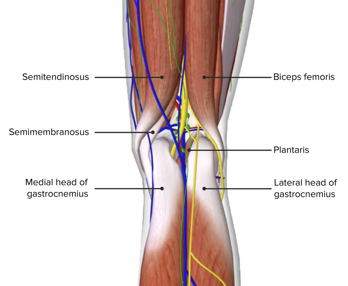


Popliteal Fossa Concise Medical Knowledge
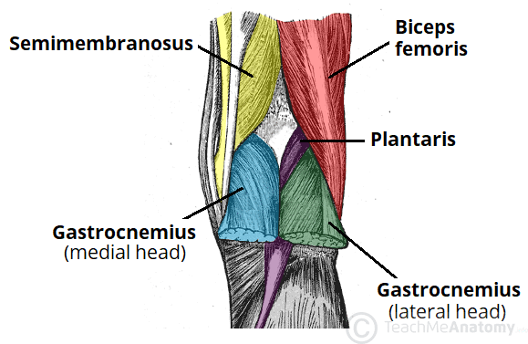


The Popliteal Fossa Borders Contents Teachmeanatomy
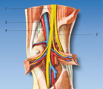


Popliteal Sciatic Nerve Block Anesthesia Key


Anatomy Of The Left Popliteal Fossa Download Scientific Diagram


Popliteal Fossa Radiology Reference Article Radiopaedia Org
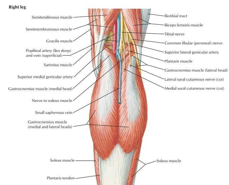


Popliteal Fossa Anatomy And Contents Bone And Spine


Poplitealfossa Explore Facebook


1
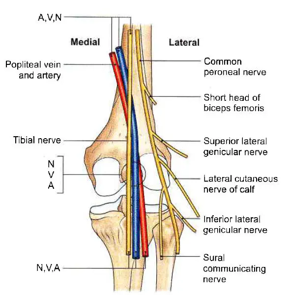


Popliteal Fossa Complete Anatomical Overview Learn From Doctor


Diagram Of Normal Popliteal Fossa Download Scientific Diagram
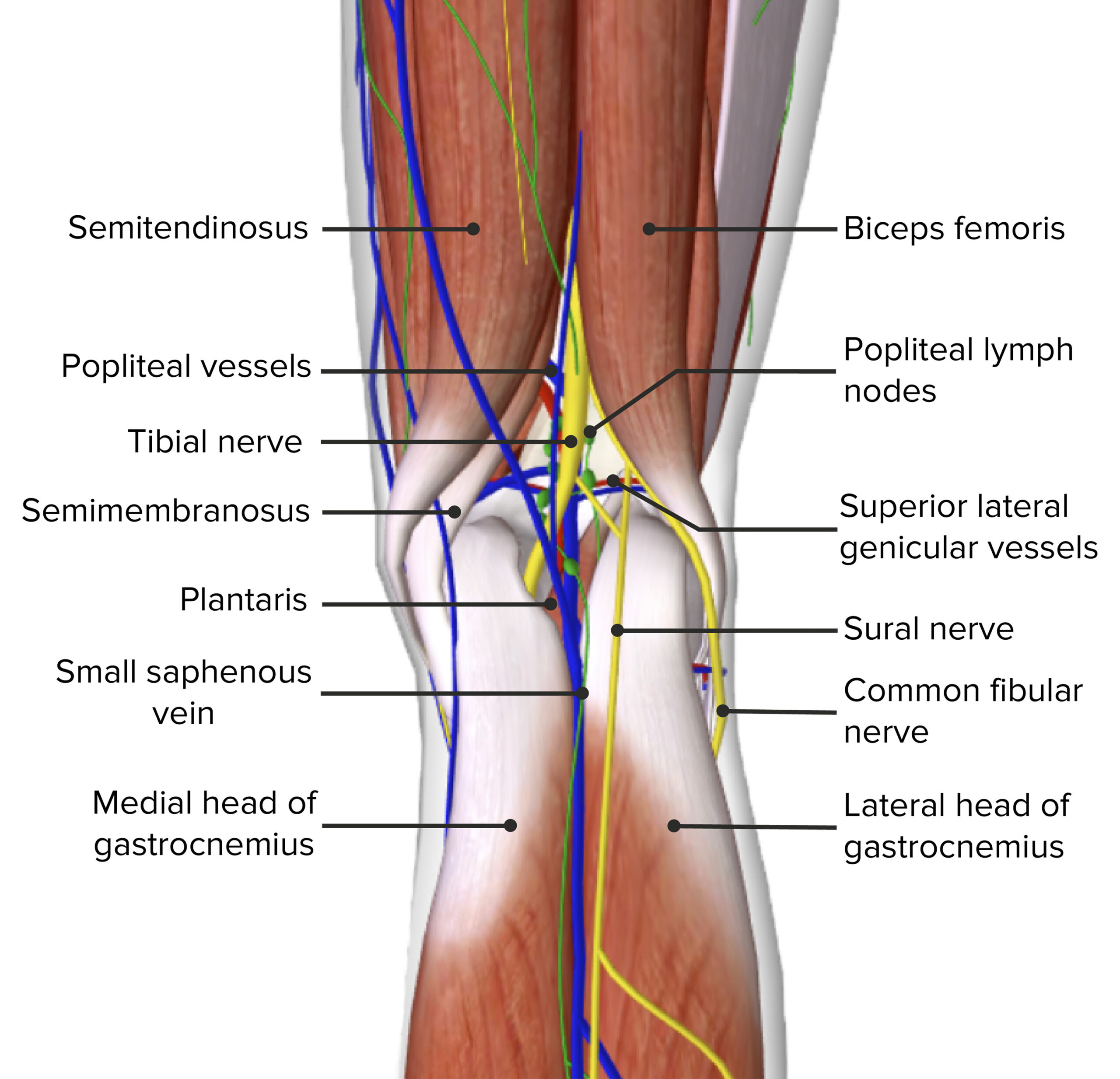


Popliteal Fossa Concise Medical Knowledge


Popliteal Fossa Boundaries Contents Anatomy Tutorial Youtube


Safe Zones For Pin Placement


Anatomy Of The Popliteal Fossa Anatomy Drawing Diagram
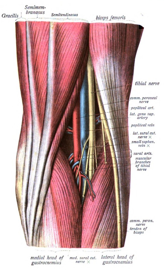


Popliteal Fossa Wikipedia


Popliteal Fossa World Wide Lifestyles Weight Loss And Gain Tips
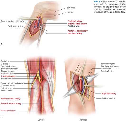


Surgical Exposure Of The Lower Extremity Arteries Thoracic Key


Anatomy Of Popliteal Fossa Anatomy Drawing Diagram


Safe Zones For Pin Placement
:watermark(/images/watermark_only.png,0,0,0):watermark(/images/logo_url.png,-10,-10,0):format(jpeg)/images/anatomy_term/vena-poplitea/1qimxsaGtAeyhFKKAmU4Qw_bzAdvntzTh_Vena_poplitea_1.png)


Popliteal Fossa Anatomy And Contents Kenhub


Dissection Of The Popliteal Fossae With The Reference Points Marked Download Scientific Diagram


Ultrasound Guided Popliteal Block Wfsa Resources


1


Safe Zones For Pin Placement


The Popliteal Fossa Boundaries Contents Youtube


Popliteus Muscle Nerve Human Leg Popliteal Fossa Popliteal Artery Angle Text Anatomy Png Pngwing


Popliteal Fossa Wikipedia


Muscles Of The Lowber Limb Fascial Compartments Of


Physioosteobook Facebook


Anatomy Of The Popliteal Fossa Everything You Need To Know Dr Nabil Ebraheim Youtube


Bats Better Anaesthesia Through Sonography
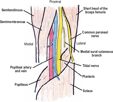


The Diagnostic Anatomy Of The Sciatic Nerve Neupsy Key
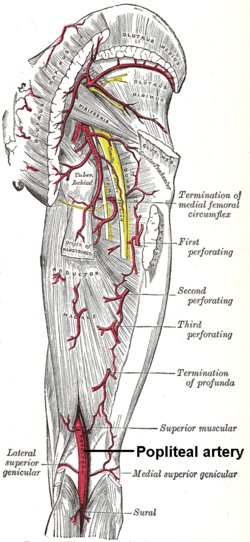


Popliteal Artery Wikipedia
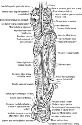


Nerves Blood Vessels And Lymphatics Of The Knee And Leg Dummies
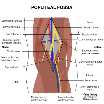


Popliteal Fossa Anatomy Mnemonic Radiology Reference Article Radiopaedia Org


Popliteal Fossa Block Regional Anesthesia Mitch Medical Healthcare
:watermark(/images/watermark_only.png,0,0,0):watermark(/images/logo_url.png,-10,-10,0):format(jpeg)/images/anatomy_term/gastrocnemius-muscle-7/EzO9qof1YSPzdJNYV4jWhQ_Gastrocnemius_muscle_magni.png)


Popliteal Fossa Anatomy And Contents Kenhub
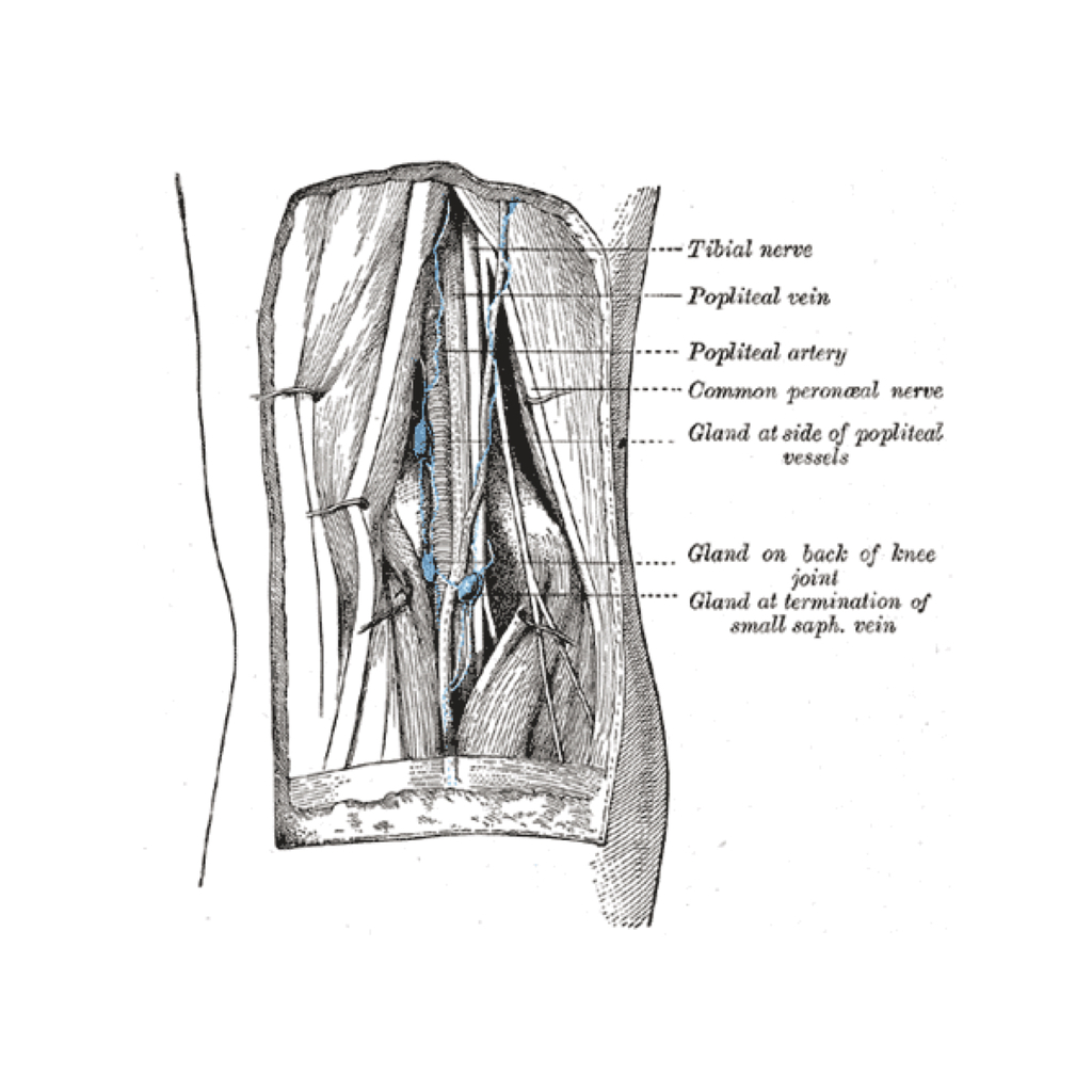


Lymphatics Of The Popliteal Fossa Gray S Illustration Radiology Case Radiopaedia Org
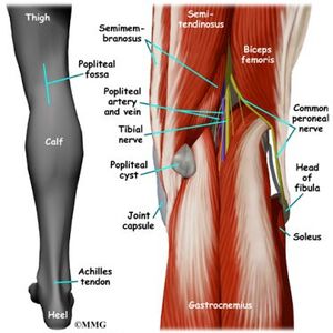


Popliteal Fossa Physiopedia


Http Ksumsc Com Download Center Archive 1st 439 2 muscloskeletal block Team work Anatomy 15 popliteal fossa 2c back of the leg and sole of the foot Pdf


Popliteal Fossa


1
:watermark(/images/watermark_only.png,0,0,0):watermark(/images/logo_url.png,-10,-10,0):format(jpeg)/images/anatomy_term/arteria-poplitea/ky8xkxCiBfMrBEsLSn2w_Arteria_poplitea_dorsal.png)


Popliteal Artery Anatomy Branches Location And Course Kenhub


Steps Of Popliteal Block Nerve Anatomy Anatomy Sciatic Nerve


Contents Of The Popliteal Fossa Anatomy Meniscal Tear Gastrocnemius Muscle


Thigh Knee And Popliteal Fossa Knowledge Amboss


Www Kgmu Org Digital Lectures Medical Anatomy Dr Kaveri Anatomy Lecture On Popliteal Fossa Pdf
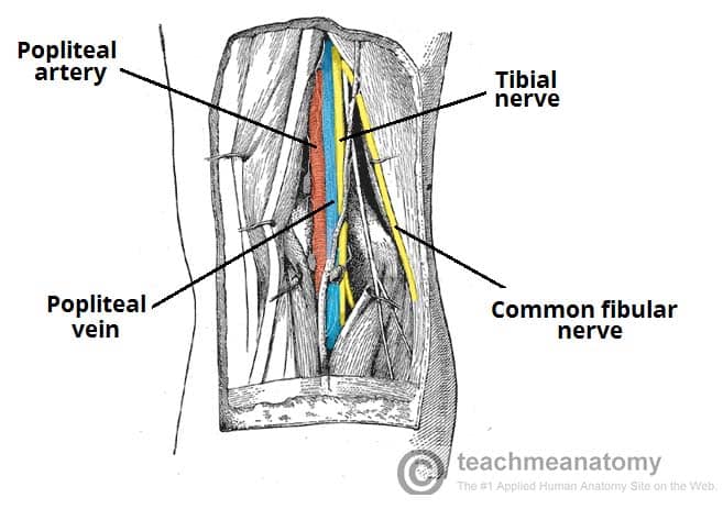


The Popliteal Fossa Borders Contents Teachmeanatomy
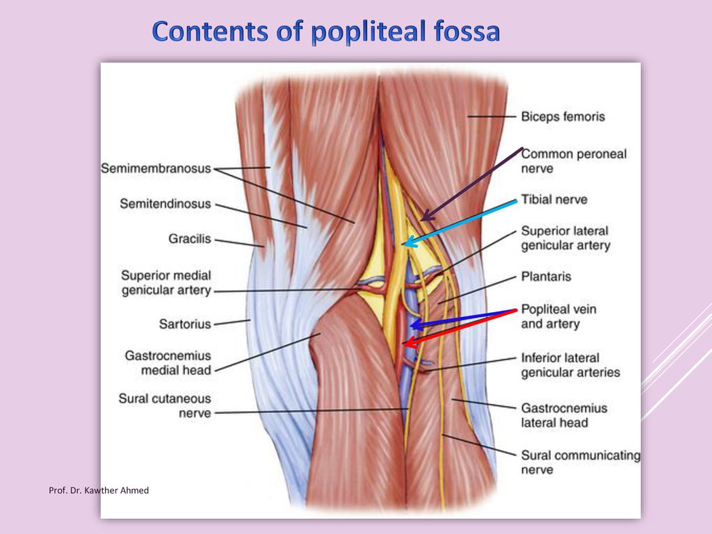


Popliteal Fossa By Prof Dr Kawther Ahmed Prof Dr Kawther Ahmed Ppt Download


The Veins Of The Popliteal Fossa The Dorsal Or Thigh Extension Of The Download Scientific Diagram


Cambridge Orthopaedics Popliteal Block Uk


1


Popliteal Artery Location Entrapment Popliteal Artery Aneurysm


Hijamah Wet Cupping Therapy For The Popliteal Fossa The Knee Pit Peshawar Hijama Center


Jaypeedigital Ebook Reader
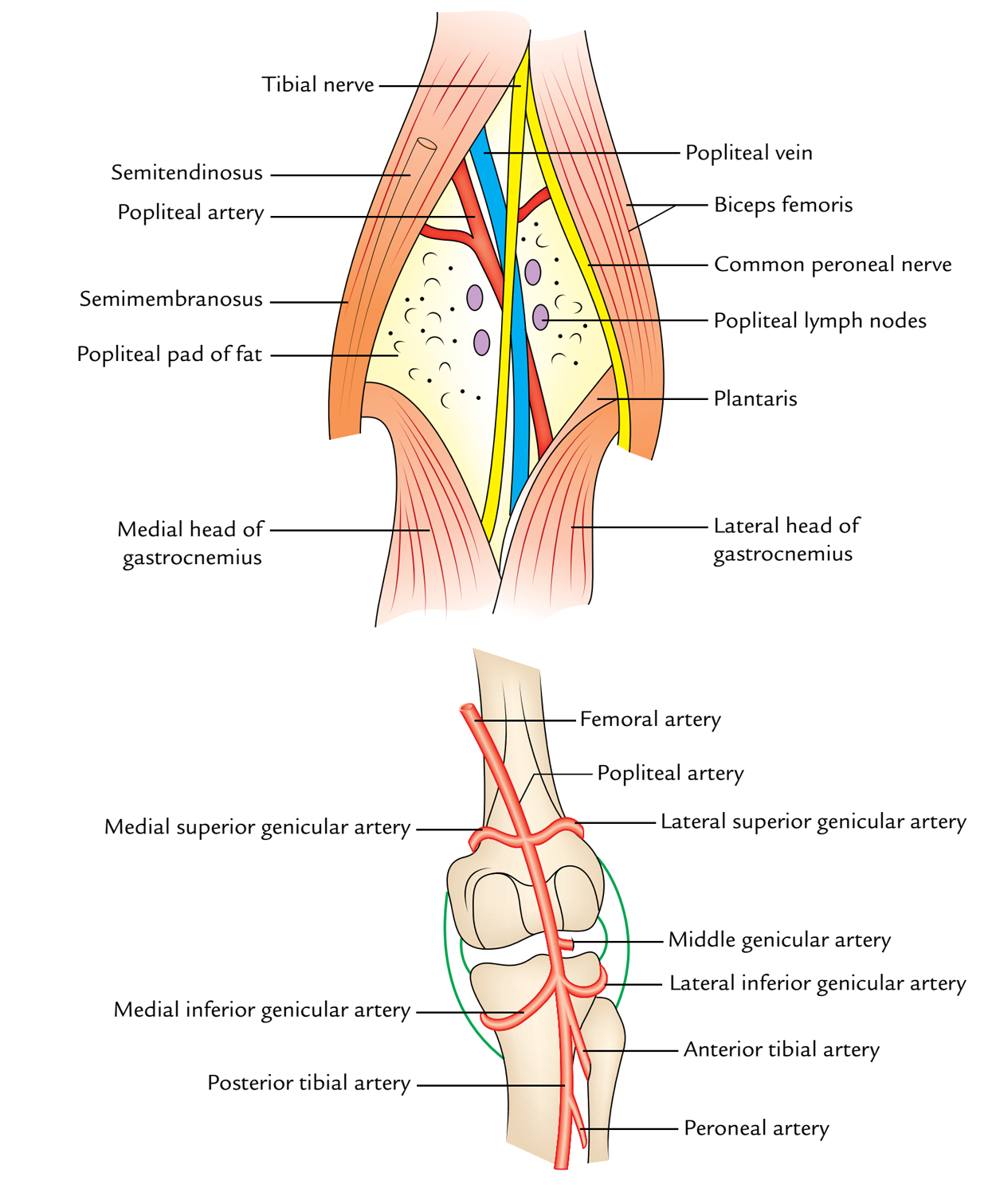


Easy Notes On Popliteal Fossa Learn In Just 4 Minutes Earth S Lab


Popliteal Fossa Bf Biceps Femoris Pa Popliteal Artery Pv Download Scientific Diagram


Http Www Kgmu Org Digital Lectures Medical Anatomy Lecture On Popliteal Fossa Pdf
/GettyImages-87313663-16bdfeaf37d048dbaef06b4f00b269b5.jpg)


Popliteal Vein Anatomy And Function
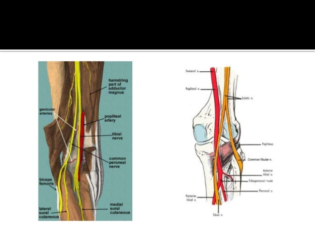


Popliteal Fossa


Popliteal Artery Entrapment Syndrome Wikipedia


Popliteal Fossa Boundaries Contents And Applied Aspects Anatomy Qa
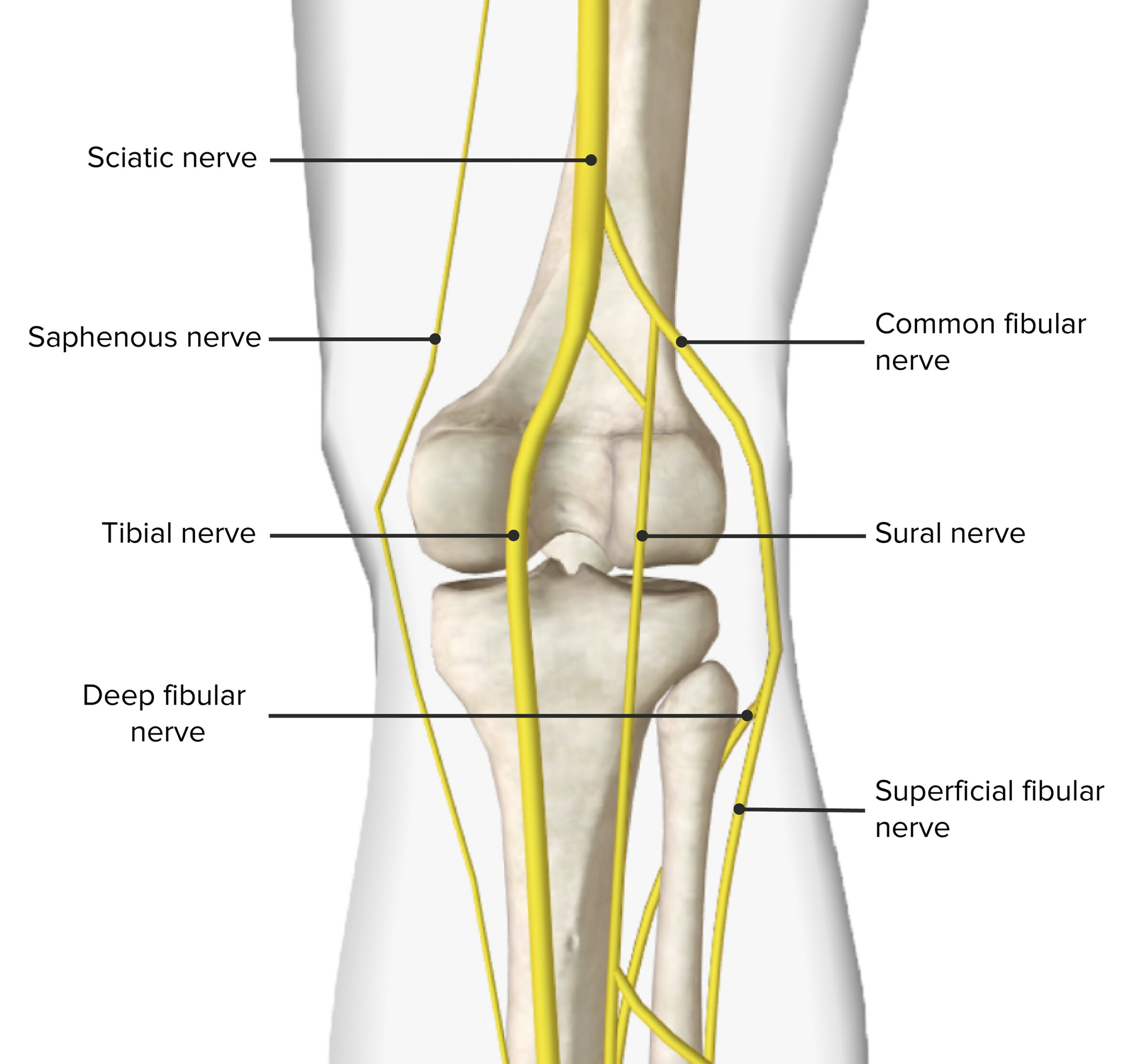


Popliteal Fossa Concise Medical Knowledge


Popliteal Fossa And Knee Joint Last S Anatomy Regional And Applied
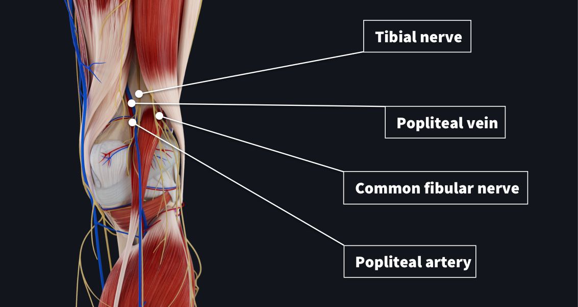


The Popliteal Fossa Complete Anatomy


3 10 Popliteal Fossa And Leg Flashcards Quizlet


No comments:
Post a Comment